Prilado y recubrimiento de dinitramida de amonio (ADN) para propulsantes sólidos “verde” en la mezcla de tolueno mediante sonicación de ultrasonido

La ADN (H4N4O4) en su forma genérica tiene una estructura acicular larga que dificulta su carga en estado sólido. Por ello, es conveniente optimizar su morfología cristalina en forma octagonal. Por otra parte, la gran higroscopia del ADN lo inutiliza en climas húmedos, Por lo que es necesario la encapsulación de sus cristales en un polímero hidrofóbico. En el trabajo de la referencia, ADN fue sintetizada por nitración de sulfamato de potasio (H2KNO3S) con mezcla sulfonítrica. El producto fue luego recubierto con un polímero (poliestireno o HTPB) y tratado con ultrasonido para obtener partículas semiesféricas recubiertas de ADN. 

1. Introduction

The utility of Ammonium Dinitramide (ADN) as a highly energetic oxidizer has several advantages in terms of performance, stability [1], and environmental friendliness as compared to conventional Ammonium Perchlorate (AP)-based propellants [2]. However, solid grain casting of raw ADN is hindered by its needle shape [3] and by its high moisture absorption tendency and lower critical humidity level (55.2% RH). Previous experiments have shown that the moisture absorption of ADN is higher than that of AP [4]. The hygroscopicity of ADN can be decreased by coating it with hydrophobic polymers [3,5,6] such as hydroxyl terminated polybutadiene (HTPB), polystyrene (PS), and polyacrylate (PA). Otherwise, specifications given on the particle size, particle layout geometry, and surface area under the experimental conditions have seldom been described in the literature. These factors, i.e., particle morphology and water absorption [7], are known to reduce the Theoretical Maximum Density (TMD) and, consequently, the specific impulse of the ADN solid grain.
Many methods have been utilized to alter the crystal particle morphology of energetic materials, such as melt prilling [6], ultrasound-assisted crystallization [7], solvent crystallization [8], and spray crystallization [9]. Melt prilling, which is an important process in conventional production of ADN, possesses several drawbacks, such as volumetric expansion in the melting phase and it being hazardous due to high operating temperature. In fact, ADN decomposes slowly in its molten condition, generating nitrous oxides [10,11] and creating cavities and fractures during solidification into the final grain. During solidification, a volumetric shrinkage of up to 14% creates foam on the surface of ADN grain [10].
The ultrasonic method is interesting as it acts as a source of mechanical energy, allowing us to reduce the ADN particle size and changing the crystal morphology. The importance of improving the crystal morphology can be highlighted by its ability to increase the specific impulse by 30% to 50% [12]. Successful modification of RDX crystal morphology by ultrasound was previously reported [9,13], but not for ADN. It is known that ultrasound treatment can induce oxidation reactions for many substrates, as reported in [14,15]. However, ADN does not undergo decomposition or oxidative reactions during such treatment, unless sodium hydroxide was added [16].
In this study, we investigate the use of an ultrasound sonication technique to alter the morphology and improve the hygroscopicity of the ADN particles. Scanning electron microscopy (SEM) is used to examine the change in morphology. From the SEM images, the change in particle shape can be observed, while the mean particle size can be measured. Water absorption tests were carried out to evaluate the effect of polymeric coating, i.e., PS and HTPB, on the hygroscopicity of the ADN particles. Finally, the thermal stability of the coated ADN particles are studied using differential scanning calorimetry (DSC).

2. Materials and Methods

2.1. ADN Synthesis

ADN used in this study was synthesized by nitration of potassium sulfamate with nitric acid and sulfuric acid at −45 °C [17,18].
First, potassium sulfamate was prepared in bulk by the neutralization of potassium hydroxide with sulfamic acid at stoichiometric ratios. The neutralized solution was concentrated to 50% volume in a rotary evaporator. It was then added into ethanol at a 1:2 ratio by volume to obtain precipitation. The precipitate of potassium sulfamate was filtered, washed with ethanol, dried overnight at 70 °C, and stored for use in further experiments.
In the second step, potassium sulfamate was nitrated with a mixture of nitric acid and sulfuric acid at −45 °C for 25 min. The nitration reaction was stopped by diluting it with crushed ice. Subsequently, the diluted mixture was neutralized by potassium hydroxide. The mixture was dried in a rotary evaporator at 60 °C. The dried powder was extracted with acetone and filtered. Then, 2-propanol was added into the acetone solution to obtain precipitation. The precipitation was filtered and discarded. The solution was evaporated in a rotary evaporator at 60 °C to half of its initial volume. n-hexane was added into the solution until the precipitate of potassium dinitramide (KDN) was visible. The precipitated KDN is usually in the range of 80 to 90 wt % purity, as determined by UV spectroscopy. The KDN was then converted to ADN by reacting it with ammonium sulfate in water.
After the conversion to ADN, the entire solution was evaporated at 70 °C to obtain dry powder. This dry powder was then extracted with ethyl acetate and collected. The collected ethyl acetate solution of ADN was then dried in a rotary evaporator to obtain pure ADN. The purity was determined by UV spectroscopy to be in range of 99 to 99.5 wt %.

2.2. Melt Prilling and Coating of Pristine ADN by Conventional Method

Melt prilled ADN samples were prepared according to the method given in [6]. Here, 0.5 g of dried pristine ADN was added in 50 mL paraffin oil, followed by the addition of 0.01 g (2 wt % w.r.t. ADN) Cab-o-sil at 100 °C under strong agitation. The mixture was cooled to room temperature. The prilled ADN was then filtered and washed with n-hexane to remove traces of paraffin oil. The filtrate product of prilled ADN particles were then added to a mixture of polystyrene (PS) in 50 mL dichloromethane and evaporated in a rotary evaporator to obtain coated AND [6]. In the second experiment, PS was replaced with hydroxyl-terminated polybutadiene (HTPB).

2.3. Prilling and PS Coating of Pristine ADN Particles by Ultrasound Sonication

Here, 0.5 g ADN particles were added to 50 mL of toluene mixture, which also included 0.01 g Cab-o-Sil (2 wt % w.r.t. ADN), 0.01 g cetyltrimethylammonium bromide (CTAB) (2 wt % w.r.t. ADN), 0.01 g graphene powder (Graphene Nano powder, 8 nm flakes AO-2, Graphene Supermarket, Calverton, NY, USA) (2 wt % w.r.t. ADN), and 0.025 g PS (5 wt % w.r.t. ADN). The mixture was then sonicated (Elmasonic P, Elma, Singen, Germany) for 60 min at 60 °C at 37 kHz in a glass test tube. After sonication, the mixture was poured into a beaker containing 100 mL of n-hexane at room temperature and again sonicated for 5 min to avoid particle agglomeration. Then, the mixture was evaporated at 45 °C under vacuum to obtain ADN particles coated with PS. The experimental setup is shown in Figure 1. The experiments were repeated by replacing toluene with dichloromethane, n-hexane and petroleum ether but were all unsuccessful due to poor dissipation of ADN particles and evaporation during sonication.
Figure 1. Experimental setup of Ammonium Dinitramide (ADN) sonication. Pristine ADN particles sonicated in toluene mixture.

2.4. Prilling and HTPB Coating of Pristine ADN Particles by Ultrasound Sonication

Toluene mixture was prepared according to the same procedures in Section 2.3without the adding of PS. An HTPB mixture, which consisted of toluene diisocyanate (TDI), glycerol, and HTPB monomer, was prepared separately. Composition of the mixture is given in Table 1. The HTPB mixture was added into the toluene mixture together with ADN particles.
Table 1. Composition of hydroxyl terminated polybutadiene (HTPB) in toluene mixture.
The mixture was then sonicated for 60 min at 60 °C at 37 kHz in a glass test tube. After sonication, the mixture was poured into a beaker containing 100 mL n-hexane at room temperature and then the mixture was evaporated at 70 °C (boiling point of n-hexane is 68 °C) under vacuum to obtain ADN particles coated with HTPB. A higher temperature of 70 °C was used to cure the HTPB.

2.5. Morphology and Thermal Stability

The coated ADN samples were kept at room temperature in a humidity-controlled chamber with 20% relative humidity (RH). During the particle shape analysis under SEM (Quanta400F, FEI, Hillsboro, OR, USA), the use of high energy radiation and high zoom levels were avoided as they may cause decomposition in the ADN samples. The thermal characteristics of the samples were analyzed by DSC (Mettler Toledo 1 STAR, Mettler Toledo, Greifensee, Switzerland) of 35 °C to 300 °C at 10 °C min−1 heating rate with a nitrogen gas purge.

2.6. Water Absorption Test

The moisture absorption tests were performed by placing 0.3 to 0.5 g of the sample in an enclosed environment with a uniform airflow rate of 5 L min−1. The humidity of the chamber was maintained at 65 ± 2% RH [20] by passing the air from a saturated solution of ammonium nitrate (AN). The weight of the sample was recorded at 60 min intervals for a total period of 240 min to determine the water absorption. The experimental setup is shown in Figure 2.
Figure 2. Experimental setup for moisture absorption test. The sample is in an enclosed container exposed to wet air and weighted periodically.

3. Results and Discussion

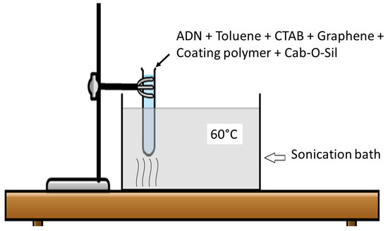
The main objective of these experiments was to increase the density ADN particles by altering the morphology of pristine ADN particles into spherical particles and to add an effective hydrophobic polymer coating. The conventional prilling method involves the melting of ADN. This could pose a potential safety hazard as the energetic ADN particles may decompose during the prilling process. Therefore, the new method based on ultrasound treatment was utilized for alteration of ADN crystal morphology and hydrophobic polymer coating. The main criterion for the ultrasound treatment of ADN was the selection of a suitable sonication medium. First, an organic solvent, which has no solubility toward ADN but has solubility toward the coating polymer, was highly desirable. Secondly, the boiling point of the solvent needed to be sufficiently high to sustain the ultrasound treatment without evaporation. In addition, the solvent needed to be removed easily by vacuum evaporation. This was necessary because excess solvent has to be removed without exposure to atmosphere.

3.1. Selection of Sonication Medium and Polymer

The reduction in particle size is dependent on the surface energy of solids (σ) [21]. The interfacial tension (γ) is the measure of interfacial energy between two immiscible fluids, or solid-fluid, given as [21]

γ12=γ1+γ22(γd1γd2)12
Equation (1) is termed the Fowkes correlation, where γ12 is the interfacial tension and γd1 and γd2 refer to dispersion forces to the surface tension by liquid 1 and liquid 2, respectively [21]. In this case, materials 1 and 2 would be the combinations of ADN, toluene, PS, and graphene with each other, as shown in Table 2.
Table 2. Interface energy of different components calculated using Equation (2).
However, the value of dispersion forces can be assumed to be 1, giving [22]

γ12(γ112γ212)2
The relation between surface tension and solubility parameter (δ) is given as [23]

δ=4.1(γ/V1/3)0.43

where V is equivalent to volume.

Furthermore, the interfacial energy is affected by crystal structure of the prilled and coated ADN. The wetting is favored if the surface energy of solid is higher than surface tension of liquid [23]. The surface tension of ADN melt (γ=89 mN/m) [21] is very high due to hydrogen bonding and polarity, compare to water =72 mN/m, while toluene has a surface tension of σ=28.4 mN/m. The surface tension of the ADN melt was used to estimate surface energy of the ADN solid.
Based on the bond breaking rule proposed in the previous literature [24], the surface tension γliquid(ADN melt) can be correlated to surface energy by the following equation:

σsolidγliquid0.713
Thus, the surface energy of ADN solid is estimated to be σADN=124.8 mN/m.
The difference in surface tension hence favors proper wetting and dispersion of ADN in toluene, which is further enhanced by elevated temperature i.e., 60 °C. The addition of cationic surfactant, CTAB, with an estimated hydrophilic-lipophilic balance (HLB) equivalent number 13+ [25], failed to disperse the ADN in solvents except toluene. The application of ultrasound sonication facilitates rapid dispersion, which increases the solid-liquid interface.
Using the value of σADN, the interaction force between different components of the system were calculated as shown below (Table 2) with data from Krevelen [23]. In general, the adding of ADN increases the interface energy with ADN-toluence combination that exhibits the highest interface energy.
The PS and graphene adhesion to ADN can be described by the work of adhesion as given by

Wadh=γs1+γs2γs1s2
where,  γs1s2=((γs1)12+(γs2)12 )12

where γs1 and γs2 are surface energy of materials being considered.

A negative value of Wadh represents poor adhesion and reversibility of the process. Wadh values for different components of the system are shown in Table 3. They are all in the positive regime. This implies a good adhesion of ADN to PS (Wadnps=144.81 mN/m). Similarly, using the interface energy of γgr=46.7 mN/m  [26], adhesion of graphene toward ADN and PS was calculated to be Wadngr=169.11mNm and Wgrps=88.57mNm, respectively, suggesting a good compatibility among them.
Table 3. Work of adhesion for different components of the system.

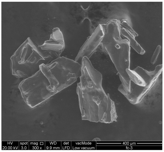
3.2. Crystal Morphology and Particle Size

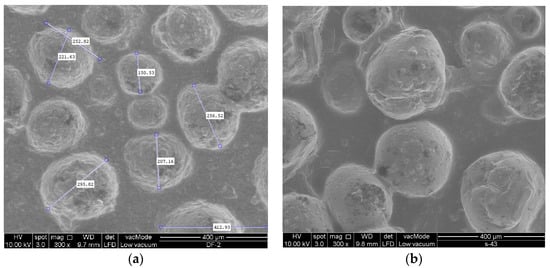
Pristine ADN synthesized in this experiment has long needle-like shape, as shown in Figure 3, compared to pristine ADN prepared by Heintz et al. [
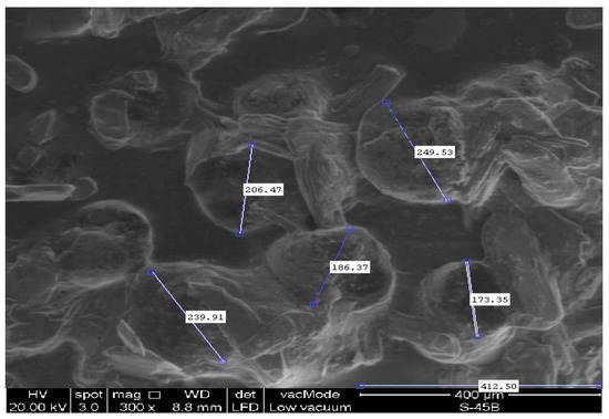
6]. Spherical ADN particles produced via ultrasound sonication followed by coating with 5% PS and HTPB are shown in Figure 4. In comparison, ADN melt prilled with conventional method and coated with 5 wt % PS is shown in Figure 5. The particle diameters were measured from the SEM images using MATLAB.
Figure 3. Scanning electron microscopy (SEM) image of pristine ADN used in these experiments.
Figure 4. (a) SEM image of ADN produced via ultrasound sonication, following by coating with 5% of PS. The particles diameter ranges from 150 µm to 295 µm; (b) SEM image of ADN sonicated and coated with HTPB. The particle diameter ranges from 155 µm to 305 µm.
Figure 5. SEM image of ADN produced by melt prilling and coating with 5 wt % polystyrene (PS).
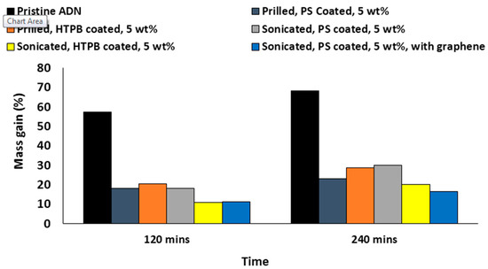
In Figure 5, the melt prilled ADN particle coating with 5 wt % PS has a size within the range of 173–250 µm. Overall, the ADN particles produced using this method have a mean diameter of 231 ± 48 µm. For ultrasound sonication, the ADN particles have a spherical shape with a particle diameter that ranges from 150 µm to 295 µm, with an overall mean diameter of 225 ± 30 µm. Although the existing ultrasonication set-up could not reduce the mean particle size of the coated ADN particles substantially, it reduced the diameter to the smallest dimension of 150 µm. This is approximately 13% smaller than its counterpart produced using the prilling method. This suggests the ultrosonication method can be optimized further. Nevertheless, the spherical shape of the coated ADN particles produced using the ultrasonication method enables a higher packing density.

3.3. Water Absorption Testing

The water absorption testing was carried out using pristine ADN, prilled ADN and sonicated ADN particles, respectively. The mass gain in the samples are shown in Figure 6. In general, the high water absorption in ADN particles is due to the presence of a hydrogen bond in its crystal structure. It is obvious from Figure 6 that the prilling and sonicated coating of ADN has reduced the water absorption as compared to pristine ADN. ADN particles sonicated coating with 5 wt % of PS achieved up to 30% in mass gain after 240 min, while sonicated coating with the identical concentration of HTPB reached only 18% of mass gain. The testing of ADN coated with hydrophobic PS embedded with graphene flakes shows a reduction in moisture absorption. This effect is particularly significant after prolonged duration, i.e., 240 min. The graphene is a hydrophobic material [26] and acts as moisture barrier because of its high aspect ratio [27,28].
Figure 6. Mass gain in pristine ADN, melt prilled, and sonicated coating of the ADN particle.

3.4. Effect of Additives

Several attempts were made to produce coated ADN without graphene, CTAB, and Cab-o-sil. In the absence of CTAB, approximately 70% of prilled ADN was still left at the bottom of the toluene mixture even after 180 min of sonication. In another attempt to produce coated ADN without graphene and Cab-o-sil, the prilled ADN particles agglomerated in the drying and evaporation steps.
CTAB consists of an anionic long carbon chain (19 carbons) and a relatively weak methylammonium bromide cation, which makes it cationic surfactant [29].
The cationic part attracts ADN because of the overall negative charge of ADN molecules. Furthermore, the anionic part of CTAB attracts methyl pendant group of toluene by virtue of partial charges leading to proper suspension of ADN in toluene.
In this work, Cab-o-sil was added as a protective colloid to promote emulsification and reduction in viscosity [30]. Its non-Newtonian behaviour provides better mechanical energy transfer to surface boundary between toluene and AND particles. In addition, it also decreases the agglomeration of particles by inducing steric hindrance [30]. In this case, the mechanical energy required to reduce the ADN particle size is provided by the ultrasound sonication.
The use of graphene in energetic materials is a rather new idea. It has only been reported for a few propellants with improvements in thermal stability [31]. However, it has been used extensively to impart hydrophobic properties in polymers, where it acts as a physical barrier or produces a lotus effect [
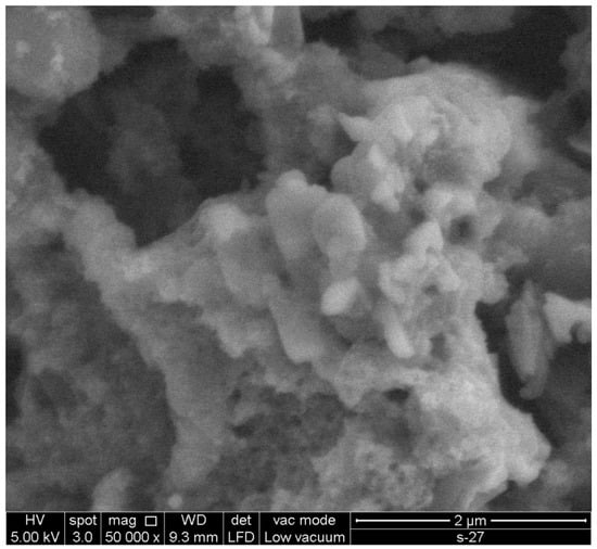
31] to reduce moisture absorption. As such, graphene was added into the ADN suspension during sonication to reduce moisture absorption. In addition, the adding of graphene is expected to reduce agglomeration of the ADN particles by increasing the distance between ADN particles. Figure 7 shows the SEM image of the agglomeration of ADN particles without the adding of graphene.
Figure 7. Agglomeration of ADN particles.

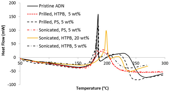
3.5. Thermal Analysis

DSC results of pristine, prilled, and sonicated ADN samples are shown in Figure 8. Pristine and prilled ADN particles were reported to have two peaks in the range of 130 to 220 °C [32,33]. The first peak around 130 °C indicates the decomposition of the dinitramide ion to produce ammonium nitrate and N2O. The second peak, which is minor peak compared to the first, indicates decomposition of ammonium nitrate [33]. All of our samples exhibit the similar trend of having two peaks. The coating of HTPB and PS on ADN particles has shifted the second peak to the range of 180 to 230 °C. Higher content of HTPB (20 wt %) causes slow burning of ADN sample. Thus, it has the first peak (at 200 °C) in higher temperature as compared to the other peaks. The trend in the DSC result for sonicated ADN particles is similar to the that of pristine and prilled ADN particles. This indicates that the technique used to coat the ADN particles has a negligible effect on the thermal properties of ADN particles. However, an exception was observed for sonicated ADN particles with 20 wt % HTPB. It has higher melting temperature due to the increased amount of HTPB, contributing to the delay in burning. The overall heat release (enthalpy) during decomposition is 1.78–3.35 kJ/g (220–416 kJ/mol), which is in good agreement with published values [34,35,36]. A summary of the results from thermal analysis is tabulated in Table 4.
Figure 8. Thermal decomposition in differential scanning calorimetry (DSC) obtained for samples at 10 °C min−1 heating rate.
Table 4. Details of thermal testing.

4. Conclusions

In this work, a method for the prilling and coating of ADN particles in an organic solvent using ultrasound sonication was demonstrated. The demonstrated method is suitable for the production of ADN particles in highly humid conditions beause ADN is not exposed to atmosphere throughout the process. The morphology of the particle has changed to semi-spherical. The particle size obtained is comparable to the melt prilling method. The combination of Cab-o-sil, CTAB, and graphene and HTPB provides good adhesion on the pristine ADN particles. The resulting hydrophobic coating has reduced the water absorption of the ADN particles. The ultrasound sonicated coating method does not change the thermal stability of the particles. This method should provide an alternative route in the production of densely packed ADN particles for use in space propulsion systems.
Acknowledgments

This work was supported by eScience Fund from Ministry of Science, Technology and Innovation Malaysia (MOSTI), Project number: 04-02-12-SF0160.
Author Contributions

All authors contributed to experimental design and data analysis. Asad Rahman performed the experiments and wrote the paper. Jitkai Chin contributed to project overview. Kean How Cheah contributed to data analysis and manuscript writing.
Conflicts of Interest

The authors declare no conflict of interest. The founding sponsors had no role in the design of the study; in the collection, analyses, or interpretation of data; in the writing of the manuscript, and in the decision to publish the results.

Fuente: http://www.mdpi.com